
FIGURE 6.
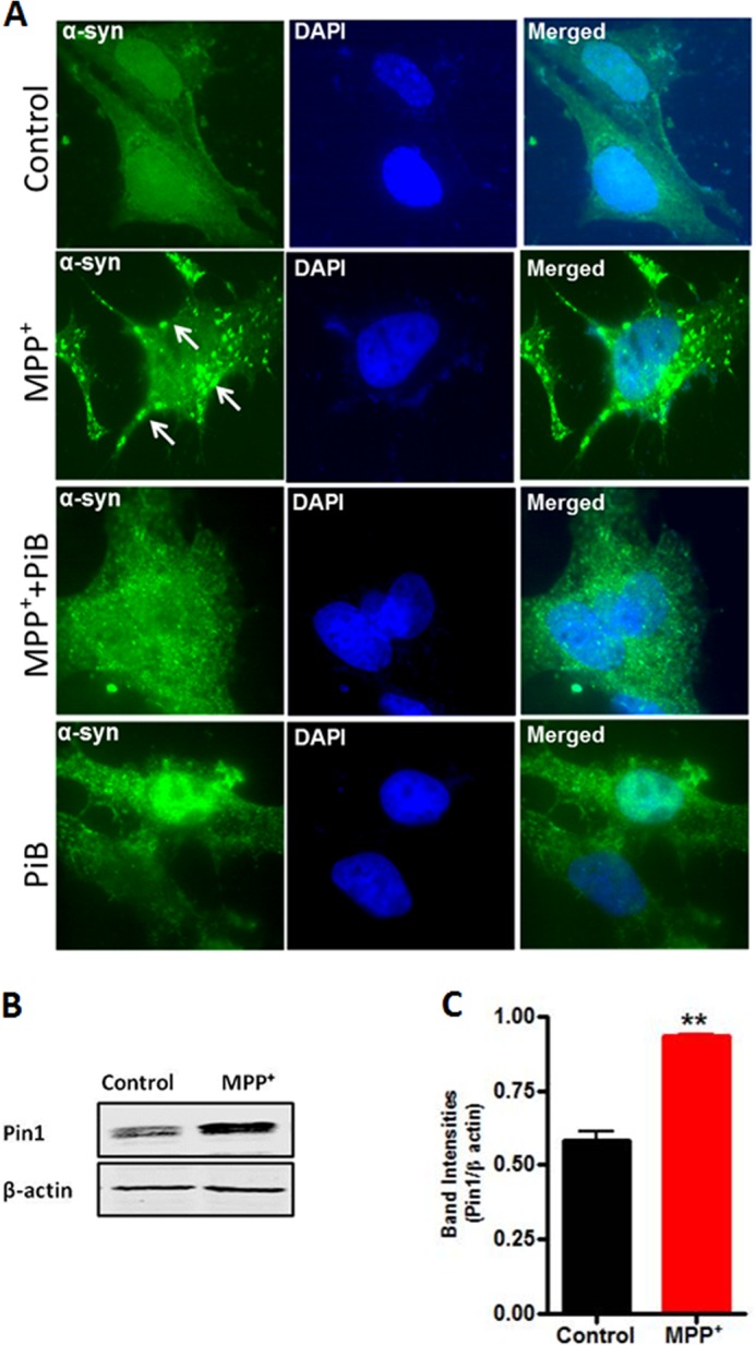
Pin1 mediates the formation of α-synuclein aggregates in response to MPP+ in N27 dopaminergic cells. A, α-synuclein stably expressing N27 cells were pretreated with the Pin1 inhibitor PiB (3 μm) for 12 h and then exposed to MPP+ (300 μm) along with PiB (3 μm) for 24 h. Cells were fixed and immunostained for α-synuclein. The nuclei were counterstained by DAPI. Images were obtained using a Nikon Eclipse T1 fluorescence microscope. Green, α-synuclein; blue, nucleus. The white arrows point to α-synuclein-positive inclusions. Magnification, 100×. Representative immunofluorescence images are shown. B, MPP+ induced Pin1 up-regulation in α-synuclein stably expressing N27 cells. Cells were treated with MPP+ for 24 h, and Pin1 expression was measured by Western blot. C, the graph represents densitometric analysis of Pin1 levels in panel B. Results are the mean ± S.E. of at least three independent experiments. **, p < 0.01 versus control.