FIGURE 8.
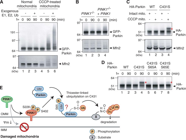
A, mitochondria collected from cells ± CCCP pretreatment were incubated at 30 °C with cytosol expressing GFP-Parkin collected from cells with intact mitochondria. In this cell-free ubiquitylation assay, CCCP-treated mitochondria stimulate autoubiquitylation of GFP-Parkin and substrate ubiquitylation toward Mfn2. B, activation of GFP-Parkin by CCCP-pretreated mitochondria depends on PINK1. Mitochondria collected from PINK1−/− MEFs following CCCP treatment do not activate GFP-Parkin in the cell-free ubiquitylation assay, whereas exogenous PINK1 complements the aforementioned defect. C, formation of an apparent ubiquitin-oxyester adduct on the Parkin C431S mutant is dependent on the presence of CCCP-pretreated mitochondria in the cell-free assay. The red asterisks indicate ubiquitin-oxyester formation unless otherwise specified. D, ubiquitin-oxyester formation of Parkin harboring C431S, C431S/S65A, or C431S/S65E mutations in the cell-free ubiquitylation experiments. The S65A mutation disrupted ubiquitin-ester formation completely, whereas the C431S/S65E mutant has weak but detectable ubiquitin-oxyester formation. E, a model for Parkin activation on damaged mitochondria. See text for details.

