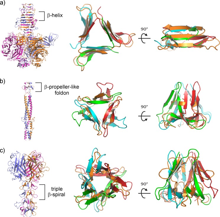
FIGURE 5.
Virus-like structural motifs observed in the Pgp3 NTD. a, endosialidase of bacteriophage K1F (PDB entry 1V0F (40)), with the β-helix region indicated (each chain shown with a different color). The bacteriophage β-helix superimposes on the N-terminal β-helix of Pgp3 shown in two orthogonal orientations. The color scheme for Pgp3 polypeptide chains is preserved as above, whereas the virus structure is colored orange for its three chains for clarity in the superpositions (two right panels). b, bacteriophage T4 fibritin with a highlighted foldon motif (PDB entry 1AA0 (46)) superimposed on the lower β-pinwheel region of Pgp3. c, adenovirus fiber with a highlighted portion of its β-spiral region (PDB entry 1Q1U (48)) superimposed on the upper β-pinwheel region of Pgp3.

