
FIGURE 10.
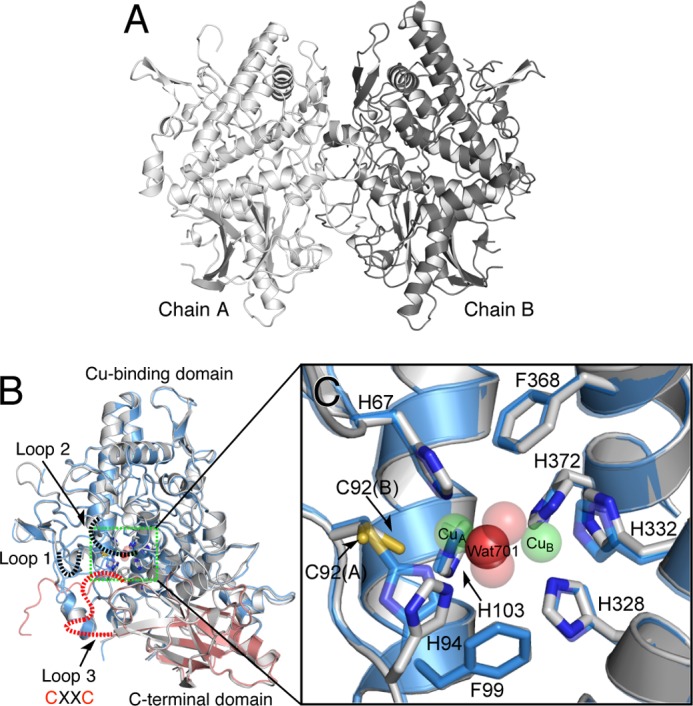
Crystal structure of melB apo-pro-tyrosinase. A, homodimeric structure of the apo-pro-form (gray and dark gray). B, subunit structures of apo-pro-form (chain A, gray) versus holo-pro-form (chain A, cyan, copper-binding domain; red, C-terminal domain). Dotted lines indicate the disordered loops colored in black and red. C, magnified view on the active site region of apo-pro-form (chain A, gray) and holo-pro-form (chain A, transparent cyan and sphere) indicated by the green rectangle. Green and red spheres indicate copper and water, respectively. Residues are shown as sticks and colored by atom type (carbon, as the respective structural element; nitrogen, blue; oxygen, red).