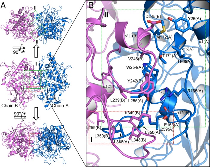
FIGURE 3.
Whole structure and dimer interfaces of melB holo-pro-tyrosinase. A, homodimeric structure in the crystallographic asymmetric unit (upper, top view; middle, side view; bottom, bottom view). Subunit interfaces of the dimeric structure have three regions at the dimer interface, as indicated by red (region I) and green (region II) rectangles. Two same interactions of region II (green) exist in the interface (upper) along a 2-fold axis. The secondary structures are colored differently according to individual chains: chain A, blue; chain B, pink. B, magnified view of the two regions of dimer interface. Residues are shown as sticks. Dotted lines indicate closet contacts between atoms.

