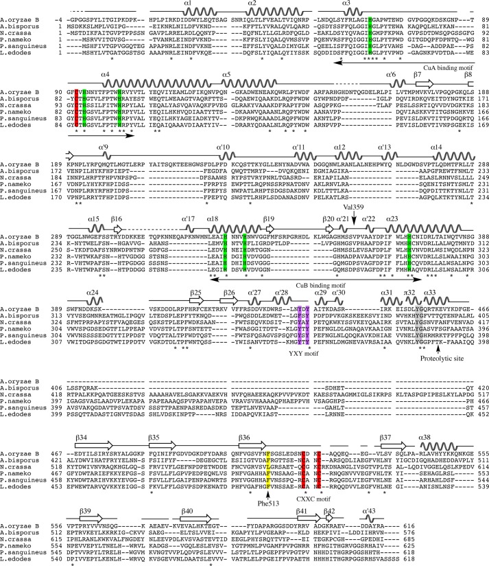
FIGURE 4.
Multiple sequence alignment of amino acid sequences of fungal tyrosinase and secondary elements of melB (α for α-helix, α′ for 310-helix, π for π-helix, and β for β-strand). The alignment was generated with ClustalW by using MEGA5 software. The conserved residues are marked with an asterisk and highlighted in rectangles (green, copper-coordinating histidines; red, conserved cysteines; yellow, the place holder; purple, YXY motif; gray, YG motif). The dashed line indicates residues not visible in the electron density. A. oryzae B, melB from A. oryzae (BD165761); A. bisporus, PPO3 from Agaricus bisporus (CAA59432); N. crassa, Neurospora crassa (CAE81941); P. nameko, Philiota nameko (AB275647); P. sanguineus, Pycnoporus sanguineus (AAX46018); L. edodes, Lentinula edodes (BAB71735).

