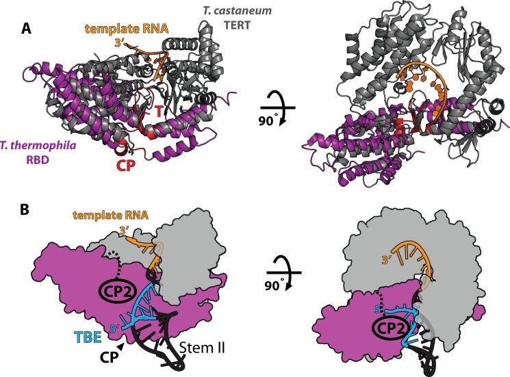
FIGURE 6.
Model of TERT-TER interactions. A, structural alignment of T. castaneum FL TERT (gray, PDB ID 3KYL) and T. thermophila RBD (purple, PDB ID 2R4G). The positions of the conserved T and CP motifs are highlighted in red. The position of the 5′ end of the template (TER residue 43) near the T motif places a constraint on the position of the TBE (residues 15–40), likely placing the TBE near the CP motif in the RBD structure. B, model of telomerase structure. The reverse transcriptase domain and C-terminal extension motifs from the T. castaneum structure are modeled in gray, and the T. thermophila RBD is in purple as in A. Based on the structural alignment in A, the CP motif likely binds the base of stem II RNA, placing stem II (PDB ID 2FRL) within the existing structural model. Our results definitively implicate the CP2 motif in stem II binding, suggesting that the CP2 motif extends toward stem II to cooperatively bind stem II with the CP motif.

