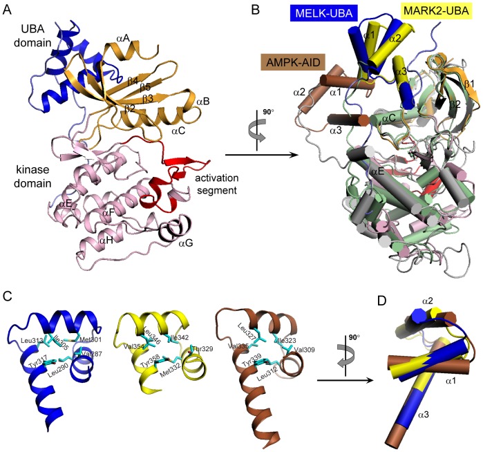
Figure 2. Overall Structure of MELK KD-UBA.
(A) Ribbon representation of MELK KD-UBA using the identical coloring scheme as in the schematic diagram in Figure 1A. All structural representations were prepared with PyMOL (http://www.pymol.org). (B) Structural comparison of MELK KD-UBA with MARK2 KD-UBA (2WZJ) and AMPK KD-AID (3H4J) upon superposition of the N-lobes of their kinase domains. The kinase and UBA domains of MARK2 are respectively shown in light green and yellow, and the corresponding domains of AMPK KD-AID in gray and brown. (C) Conserved hydrophobic cores of MELK-UBA, MARK-UBA and AMPK-AID. The hydrophobic residues are highlighted as cyan sticks. (D) Difference in the helix arrangement of three UBA domains from MELK, MARK and AMPK upon superposition of their α3 helices.

