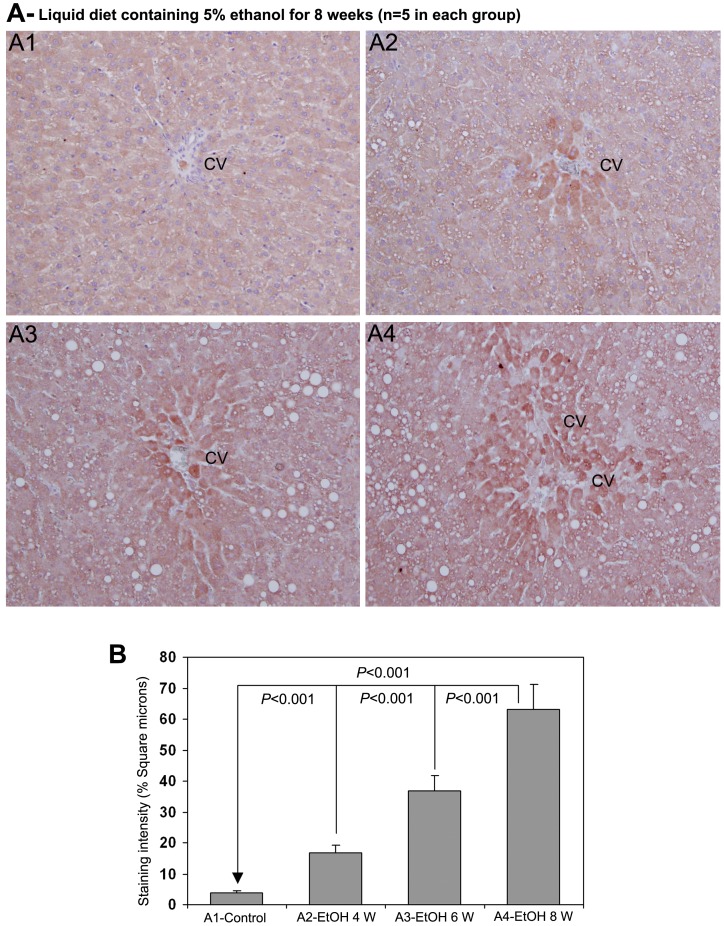
Figure 6. Immunohistochemical staining for 4-HNE in rat liver during chronic administration of ethanol (x100).
(A1) Staining for 4-HNE was completely absent in rats received control liquid diet. (A2), (A3) & (A4) Animals received liquid diet containing 5% ethanol for 4, 6 and 8 weeks, respectively. Marked staining for 4-HNE in perivenular areas in increasing order from 4−8 weeks. CV−central vein. (B) Quantitative representation of the staining intensity of 4-HNE in A1−A4 stained sections (Mean ± S.D., n = 5).

