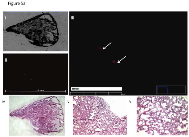
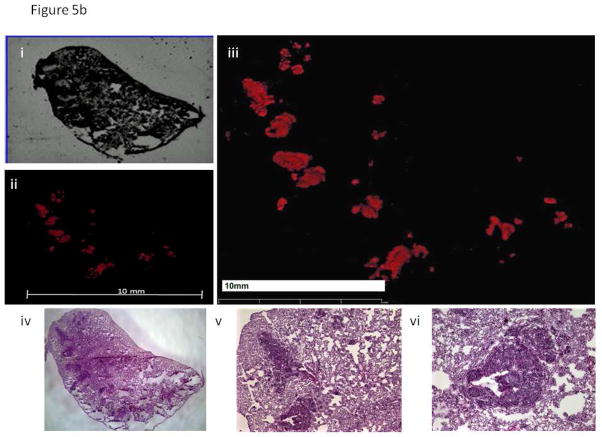
Figure 5. Histology Images of Lung Samples.
a. Representative lung sample from a mouse implanted with BxPC-3 RFP P0, small microscopic metastatic lesions are noted.
- Bright-field image of lung section from a mouse implanted with BxPC-3 RFP P0.
- Fluorescence image of lung section from a mouse implanted with BxPC-3 RFP P0.
- Enlarged fluorescence image of lung section from a mouse implanted with BxPC-3 RFP P0. Arrows indicate two small areas of metastatic foci within a section of lung tissue from a mouse harboring the BxPC-3 RFP parental line. Red fluorescence indicates the presence of tumor within the tissue sample.
- H&E image of lung section from a mouse implanted with BxPC-3 RFP P0.
- 4X magnification of H&E image of lung section from a mouse implanted with BxPC-3 RFP P0.
- 10X magnification of H&E image of lung section from a mouse implanted with BxPC-3 RFP P0.
b. Mice implanted with BxPC-3 RFP P6 had significantly more metastatic lesions to the lung than mice implanted with BxPC-3 RFP P0.
- Bright field image of lung section from a mouse implanted with BxPC-3 RFP P6.
- Fluorescence image of lung section from a mouse implanted with BxPC-3 RFP P6.
- Enlarged fluorescence image of lung section from a mouse implanted with BxPC-3 RFP P6. The red fluorescence indicates the presence of metastatic foci within the tissue sample. The area of fluorescence is significantly greater in the section of lung metastasis from a mouse harboring the BxPC-3 RFP P6 line.
- H&E image of lung section from a mouse implanted with BxPC-3 RFP P6.
- 4X magnification of H&E image of lung section from a mouse implanted with BxPC-3 RFP P6.
- 10X magnification of H&E image of lung section from a mouse implanted with BxPC-3 RFP P6.


