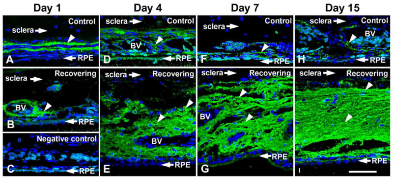
Figure 8.

Confocal images of RALDH2 protein (green) in choroids of control and recovering chick eyes following immunolabeling with anti-RALDH2 antibodies. A. robust labeling for RALDH2 is detected in ovoid cells in the stroma of choroids from a recovering eye. B. Minimal RALDH2 protein is detected in choroids of contralateral control eyes. C. Negative control section of a recovering eye (24 hours), where non-immune rabbit IgG was used in the first incubation, followed by incubation in AlexaFluor 488-conjugated goat anti-rabbit immunoglobulin. Nuclei were counterstained with DAPI (blue). From:
Summers Rada, J.A., Hollaway, L.Y., Li, N., Napoli, J., Identification of RALDH2 as a Visually Regulated Retinoic Acid Synthesizing Enzyme in the Chick Choroid. Invest Ophthalmol Vis Sci, 53:1649-1662 2012. Reproduced with permission © Association for Research in Vision and Ophthalmology.
