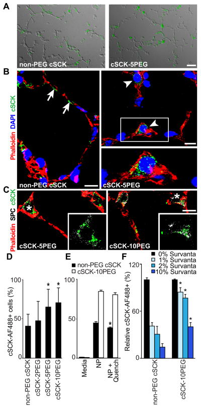
Figure 4. PEGylation of cSCKs enhances the uptake in alveolar type II epithelial cells.
(A) Fluorescence photomicrographs with differential interference contrast (DIC) overlay show the distribution of fluorescent labeled cSCK in the lung alveoli. Bar=20 μm. (B, C) Confocal microscopy of cSCK in alveolar epithelial cells stained with actin marker (phalloidin, red). (B) Non-PEG cSCKs associated with the cell surface (arrows), cSCK-5PEG internalized in alveolar epithelial type II cells (arrowheads) and DNA stained with TO-PRO-3 (blue). (C) PEGylated cSCK show internalization in type II cells immunostained for prosurfactant protein C (SPC; pseudocolored white). Insets are detail of the indicated regions (*) without phalloidin stain. Bars = 10 μm in B, C. (D–F) Uptake of cSCK in MLE 12 cells detected by flow cytometry 1 h post-incubation. (D) Quantification of cSCK uptake. n=6 independent experiments with duplicate samples (E) Quenching analysis of non-PEGylated cSCK compared to PEGylated-cSCK. Cells were incubated with the indicated cSCK form, analyzed in the absence and then in the presence anti-AF488 antibody. (F) cSCK cell uptake after incubation with artificial surfactant (Survanta) relative to media only. (E, F; n=2 independent experiments with duplicate samples). All data are the mean ± S.D. A significant difference compared to non-PEG cSCK is indicated (*, P < 0.05).

