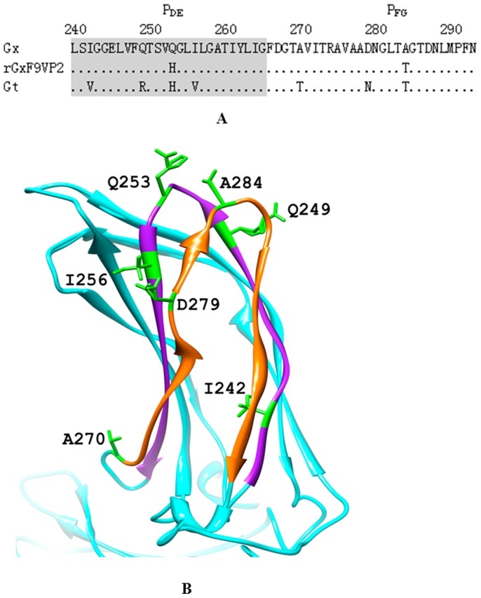
Figure 2. Comparison of the amino acid sequences of the PDE and PFG domain of IBDV VP2.
(A) Amino acid sequences of the PDE (aa 240 to 265) and PFG (aa 270 to 293) domains of VP2 of vvIBDV Gx (GenBank Accession No. AY444873), the rescued virulent strain rGx-F9VP2, and the attenuated strain Gt (GenBank Accession No. DQ403248) were compared. The PDE and PFG domains are marked, respectively. (B) The three-dimensional structure of VP2 of the Gx strain was built by the SWISS-MODEL workspace (http://swissmodel.expasy.org/workspace) and depicted using UCSF Chimera 1.6.1 (http://www.cgl.ucsf.edu/chimera); only the projection domain is shown here. PDE (purple) and PFG (orange) domains are highlighted with different color. The different residue sites of the PDE and PFG domains among these strains are highlighted with green color: I242, Q249, Q253, I256, A270, D279, A284.

