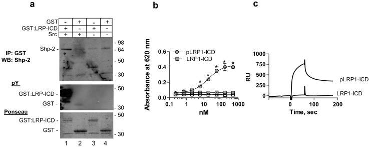
Figure 1. Phosphorylated LRP1-ICD interacts with SHP-2 with high affinity.
(a) Cell lysates from NRK fibroblasts were incubated with phosphorylated GST:LRP1-ICD (lane 1), unphosphorylated GST:LRP1-ICD (lane 3), GST and src (lane 2) or GST alone (lane 4) all bound to Glutathione-Sepharose. Following incubation and washing, eluted proteins were separated by SDS-PAGE and analyzed by immunoblot analysis for SHP-2 (upper panel), for tyrosine phosphorylation (middle panel) and for total protein by Ponseau stain (lower panel). (b) Increasing concentrations of phosphorylated (circles) or unphosporylated (squares) GST:LRP1-ICD were incubated with microtiter wells coated with SHP-2 (closed symbols) or BSA (open symbols). Bound GST:LRP1-ICD was detected with anti-GST antibodies. Curve shows the best fit to a single class of sites using non-linear regression analysis. *, absorbance values for pLRP1-ICD are significantly different from those of LRP1-ICD (p<0.0001, Students t test) (c) SPR analysis confirms binding of SHP-2 (1 µM) to immobilized phosphorylated GST:LRP1-ICD but not to immobilized unphosphorylated GST:LRP1-ICD.

