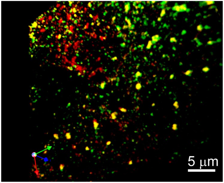
Figure 7. Co-localization of LRP1 and phospho-LRP1 in WI-38 fibroblasts.
Three-dimensional reconstruction from a stack of optical sections of the leading edge of a cell depicted in Fig. 6 (box 2). Image is rendered to show sites of colocalization facing forward. Stacks of 5 images 0.2 µm apart were captured with a BioRad confocal (Zeiss) microscope using 100X oil immersion objective; 3D reconstruction was done using Volocity (Improvision) software. LRP1 (green) and phospho-SHP-2 (red).

