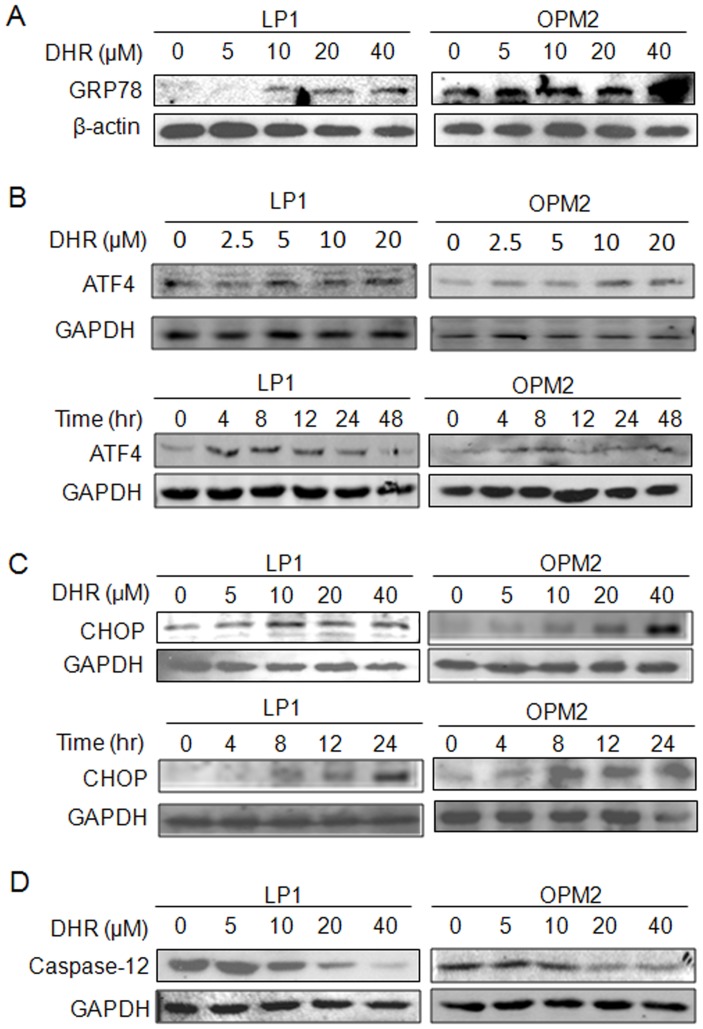
Figure 5. DHR activates the ER stress signaling in human plasma cells.
A, LP1 and OPM2 cells were treated with DHR for 24 h, the expression of GRP78 was then detected by immunoblotting. (B) and (C) LP1 and OPM2 cells were treated DHR for different concentrations and time periods before subject to ATF4 and CHOP analysis. (D) ER-associated caspase-12 was evaluated after exposed to DHR at indicated concentrations for 24 h.

