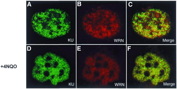
Figure 6.
Nuclear localization of Ku and WRN by immunofluorescence. Actively growing SV40-transformed GM00637 cells were untreated (A–C) or incubated with 4NQO (D–F), then fixed and subsequently incubated simultaneously with rabbit anti-WRN and goat anti-Ku as described in Materials and Methods. Anti-rabbit Texas Red-labeled and anti-goat Alexa 588-labeled secondary antibodies were used to visualize WRN and Ku by confocal microscopy at 568 and 488 nm, respectively. The images presented here depict a representative cell from each of the non-treated and 4NQO-treated cultures. In the merged images (C and F) a yellow color appears where WRN (red) and Ku (green) fluorescence signals coincide.

