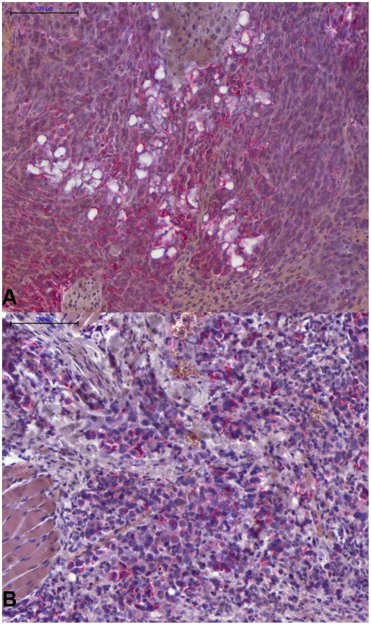
Figure 3. Human CEL and CML cells can be clearly detected by immunohistochemistry in the tissues of the scid mice.
A: Tissue section of an EOL-1 chloroma of selectin wt scid mouse. Immunohistochemical staining for human HLA-DR in red. B: Tissue section of a K562 chloroma of a wt scid mouse. Immunohistochemical staining for human mitochondria in red.

