
Figure 3. Time-dependent dissociation and association of NM-2B from filamentous α- and γ-actin.
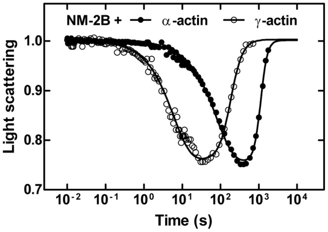
The light scattering signal normalized to the initial value shows the time-dependent dissociation and association of the actomyosin complex formed by NM-2B and α- or γ-actin. The initial drop in the light scattering signal follows the release of ATP from caged-ATP by a flash of UV-laser. The single exponential reduction in scattering intensity monitors the ATP-induced dissociation of myosin from F-actin. The following restoration of the light scattering signal is caused by rebinding of myosin to the actin filament after ATP hydrolysis is completed. As shown for NM-2B, γ-actin causes faster dissociation and association compared to α-actin. Note the logarithmic time scale on the graph.