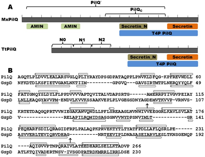
Figure 3. Conserved domains of PilQ.
Panel A. Conserved domains of MxPilQ (901 residues) and TtPilQ (757 residues) [58]. The conserved feature are drawn to scale and areindicated by shaded letters below both protein. Each has the PilQ region at their C-terminus consisting of the highly conserved Secretin domain and the region immediately N-terminal of secretin (Secretin_N). Two AMIN domains [59] are found at the N-terminus of MxPilQ but not TtPilQ. The brackets and the labels above indicate the different Y2H constructs and/or subdomains in each protein. Panel B. Structural alignment of the N-termini of TtPilQ and the T2SS secretin GspD from Enterotoxigenic E. coli (ETEC) [50], [55]. The boundaries between N0 and N1 as well as N1 and N2 subdomains in GspD are indicated by arrows (↑). The secondary structure of GspD from crystallography and that of TtPilQ predicted from modeling are indicated below and above the aligned sequences, respectively, with β strands represented by block arrows and α helices by cylinders.

