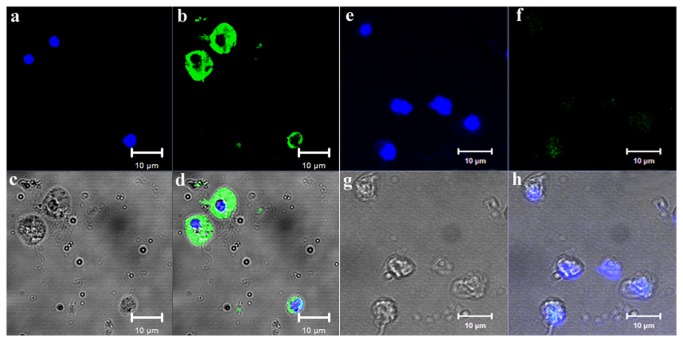
Figure 7. Localization of CfNOS protein in scallop haemocytes.
Binding of antibody to CfNOS was visualized by Alexa 488-labeled secondary antibody (green), and the nucleus of haemocytes was stained with DAPI (blue). a–d: rat polyclonal antiserum to rCfNOS, bar = 10 µm; e-h: non-immune rat serum, bar = 10 µm.

