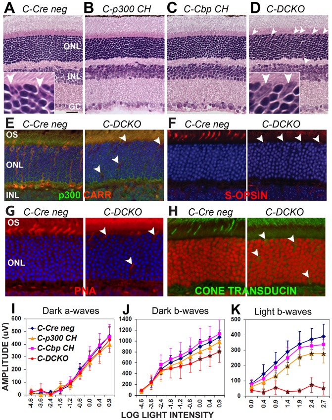
Figure 6. Ep300/Cbp conditional knockout in cones also disrupts cone structure, gene expression and function.
Cone opsin-driven Cre (CCre) was used to knock out Ep300/Cbp in cone photoreceptors; morphology, cone gene expression/distribution, and ERG function were assessed at 6–7 weeks of age. A–D. Compared to Cre negative controls (Panel A; inset shows two presumptive cones), H&E staining of CCre conditional knockout retinas reveals no major abnormalities (panels B–D), but cells with large nuclei can be seen scattered throughout the outer retina in C-DCKO mice (Panel D arrowheads and high-magnification inset). E. Cone arrestin (red) and p300 (green) expression are decreased in these cells (arrowheads). F. S-opsin expression (red) is also decreased in these cells (arrowheads), which lack outer segments. G. Peanut agglutinin labelling (red) identifies the displaced, abnormal cells in the outer retina (arrowheads) as cones. Blue in Panels E–G is DAPI labelling of nuclei. H. Cone α-transducin (green) is decreased and mislocalized to the cell bodies. Draq5 (red) marks nuclei. I. ERGs performed on 6 week old CCre mice confirmed decreases in cone-driven responses in C-DCKO retinas (red lines). Two-way repeated measures ANOVA indicated significance at p<0.0001 for dark-adapted and light-adapted b-waves. Asterisks (*) indicate significance differences (p<0.001) from Cre negative controls in post-hoc tests.

