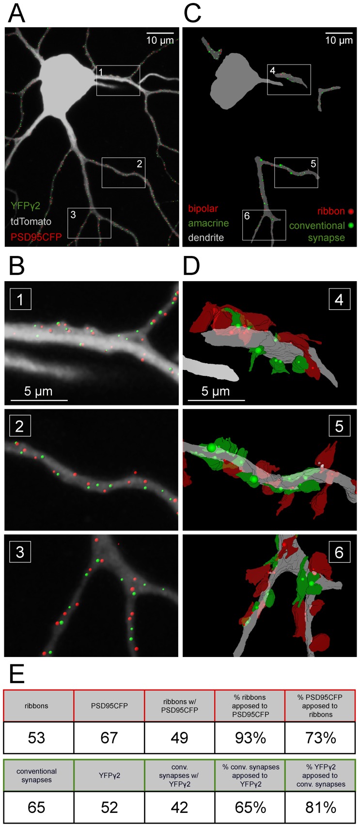
Figure 9. PSD95CFP and YFPγ2 puncta on RGC dendrites correspond to sites of bipolar cell and amacrine cell synapses.
(A) ON A-type RGC (gray) with identified PSD95 (red dots) and YFPγ2 (green dots). (B) Higher magnification of the boxed regions 1–3 in (A). (C) Three dimensional reconstructions of serial EM sections showing the locations of amacrine (conventional) and bipolar cell (ribbon) synapses on the reconstructed dendrites (gray). (D) Higher magnification of the boxed regions in (C) showing amacrine (red) and bipolar (green) cell processes apposed to the dendrite. Locations of synapses are marked by the colored dots (red, ribbon; green, conventional). The size of the dot is proportional to the length of the postsynaptic density at the synapse. (E) Table representing the number and correlation of fluorescence and anatomical profiles from the reconstructions.

