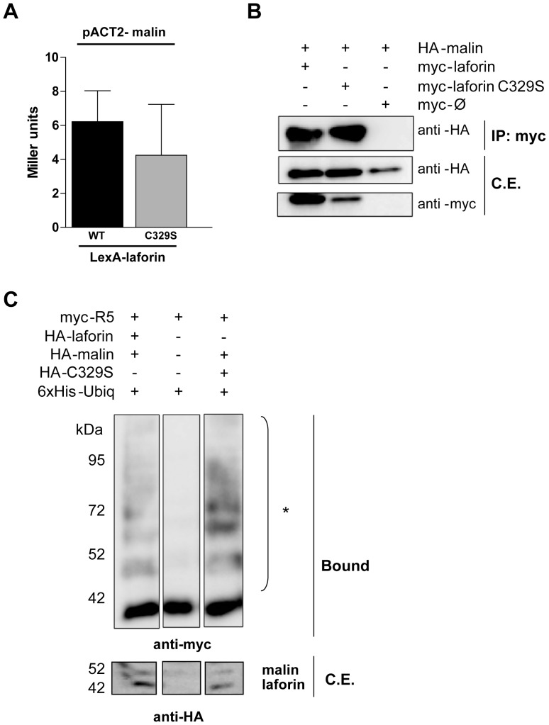
Figure 5. Laforin-C329S and malin physically interact and form a functional complex in mammalian cells.
(A) Yeast two-hybrid analysis. THY-AP4 yeast strain was transformed with pACT2-malin and LexA-laforin (WT or C329S) and the interaction was assessed by measuring the β-galactosidase activity. (B) Co-immunoprecipitation assay. HEK293 cells were co-transfected with plasmids myc-laforin (WT or C329S) and HA-malin. Cells were lysed and total lysates were incubated with anti-myc antibody and protein A/G beads. After washing, beads were boiled in loading buffer and purified proteins analyzed by SDS-PAGE and Western blot using anti-myc or anti-HA antibodies. (C) Ubiquitination analysis of R5/PTG by the laforin-malin complex. Overexpression of 6xHis-ubiquitin, pCMV-HA-malin, pCMV-myc-R5/PTG and pCMV-HA-laforin (wild type or C329S) in HEK293 cells, followed by lysis in presence of guanidinium chloride and purification of the ubiquitinated proteins by affinity chromatography using a cobalt resin. The result of the purification was analyzed using Western blot with anti-myc antibodies. Bound: proteins retained in the resin; crude extracts (50 µgr, C.E.) were immunodetected with anti-HA antibodies. *: polyubiquitinated forms.

