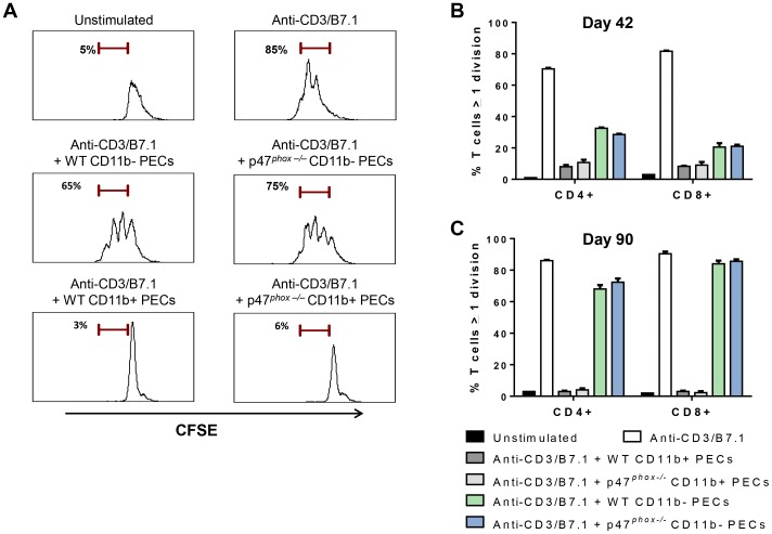
Figure 6. Myeloid peritoneal cells in MOSEC-bearing mice suppress T cell proliferation independently of NADPH oxidase.
Purified myeloid (CD11b+) PECs from MOSEC-bearing WT and p47phox−/− mice were co-cultured with CFSE-labeled splenocytes from naïve mice (E∶T ratio: 1∶1) in anti-CD3/B7.1-coated plates. After 72 h of culture, CD4+ and CD8+ T cell proliferation was assessed based on CFSE dilution as described in methods. A) Representation histograms showing that CD11b+, but not CD11b−, PECs from MOSEC-bearing WT and p47phox−/− mice (day 90) completely suppress anti-CD3/B7.1-stimulated CD4+ T cells proliferation. Anti-CD3/B7.1 stimulated and unstimulated CD4+ T cells are used as positive and negative controls, respectively. B) CD11b-enriched PECs from MOSEC-bearing WT and p47phox−/− mice at day 42 equally suppress both CD4+ and CD8+ T cell proliferation. In contrast, CD11b− PECs from the same mice incompletely suppressed anti-CD3/B7.1-stimulated T cell proliferation. C) CD11b-enriched PECs from MOSEC-bearing WT and p47phox−/− mice at day 90 completely suppressed T cell proliferation while the CD11b-negative fraction had no significant effect on T cell proliferation. Myeloid PECs collected on day 42 were pooled because of limited cell number, while non-pooled PECs from individual mice were analyzed from day 90 harvests. Individual experiments were performed using PECs from 3 mice per genotype per time point, and each experiment was repeated at least once using PECs from different mice, with similar results. Comparison between genotypes: p = NS.

