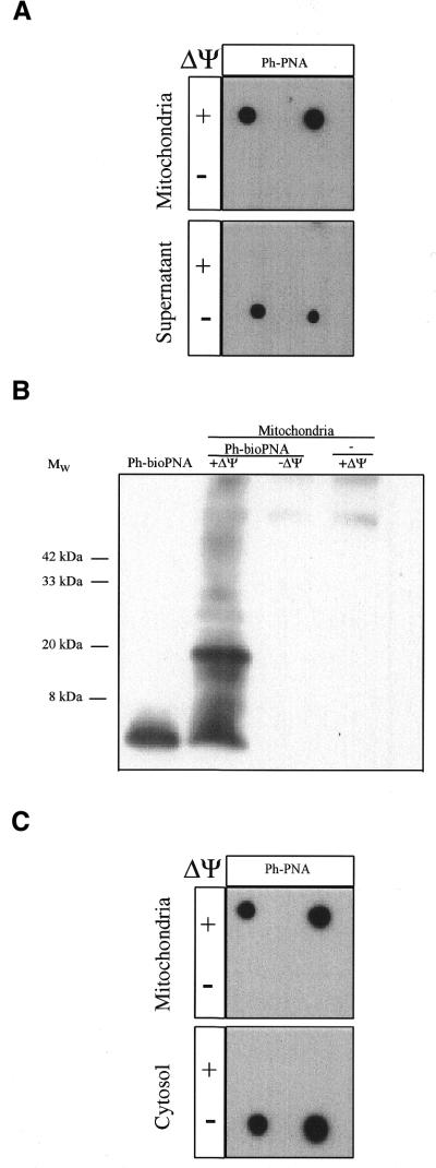
Figure 4.
Uptake of ph–PNA conjugates by mitochondria. (A) Rat liver mitochondria were incubated with 1 µM ph–PNA in the presence or absence of ΔΨm, (±10 µM FCCP). After incubation the mitochondria were pelleted through oil and the supernatant and mitochondrial fractions were probed with antitriphenylphosphonium serum. (B) A mitochondrial matrix fraction enriched for the nucleoid was isolated from mitochondria which had been incubated ±1 µM bioPNA in the presence or absence of ΔΨm, and then crosslinked with 1% formaldehyde. Samples were separated by Tris–tricine PAGE, transferred to nitrocellulose and probed for biotin. A ph–bioPNA (∼5 nmol) control was also analysed. (C) 143B cells were incubated with 1 µM ph–PNA in the presence or absence of ΔΨm, fractionated with digitionin and separated into mitochondria- and cytoplasm-enriched fractions by centrifugation through oil. Samples from both fractions were then assayed for triphenylphosphonium as above. There was no immuno-reactivity with preimmune serum in these experiments.

