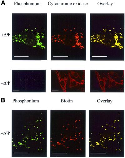
Figure 5.
Mitochondrial localisation of ph–PNA conjugates within fibroblasts by confocal immunofluorescent microscopy. Fibroblasts were incubated with 1 µM ph–PNA (A) or 1 µM ph–bioPNA (B) for 4 h at 37°C, in the presence or absence of ΔΨm. (A) Cells were fixed, incubated with antiserum against triphenylphosphonium (green) and a monoclonal antibody against cytochrome oxidase (red). In the overlaid images yellow indicates co-localisation of triphenylphosphonium and cytochrome oxidase. (B) Cells were fixed and labelled for triphenylphosphonium (green) and biotin (red). In the overlaid images yellow indicates colocalisation of the triphenylphosphonium and biotin. There was no immuno reactivity with preimmune serum in these experiments. Magnification, 1400× (B, A + ΔΨ), 600× (A – ΔΨ); scale bar, 20 µm.

