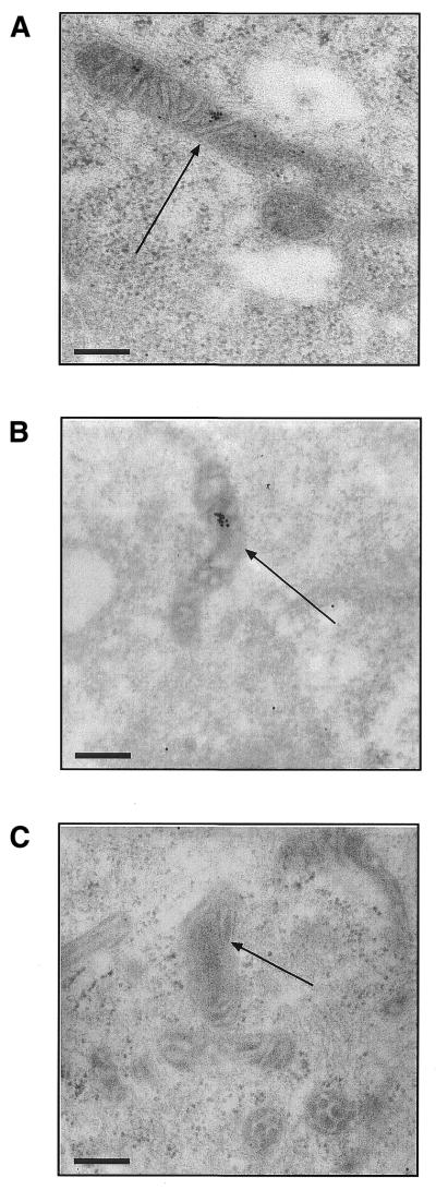
Figure 6.
Immunogold labelling of ph–PNA conjugates within the mitochondrial matrix. (A) Human fibroblasts were incubated with 1 µM ph–PNA, fixed and the intracellular localisation of triphenylphosphonium detected by immunogold electron microscopy (black dots). (B) Human fibroblasts were incubated with 1 µM ph–bioPNA and the location of the biotin determined by immunogold electron microscopy. (C) Cells were incubated with ph–PNA but the incubation with triphenylphosphonium serum was omitted, while that with the gold-linked antibody was not. Identical electron micrographs were obtained by omitting the ph–PNA conjugates from the incubation. Magnification, 52 000×; scale bar, 200 nm; arrows indicate mitochondria.

