
Fig. 2.
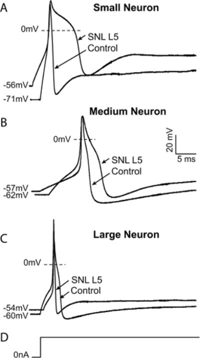
Exemplary traces showing effects of injury on action potential dimensions in control neurons and after ligation and axotomy of the fifth lumbar spinal nerve (SNL L5). (A) A typical small neuron displays prolonged action potential duration and a more depolarized resting membrane potential after injury. (B) A medium neuron exhibits prolonged action potential as well as decreased afterhyperpolarization amplitude and accelerated recovery of the afterhyperpolarization after spinal nerve ligation. (C) A large neuron develops decreased afterhyperpolarization amplitude and duration. (D) The stimulus in each case is a sustained current pulse of an amplitude just adequate for action potential generation. Scale bars apply to all traces.