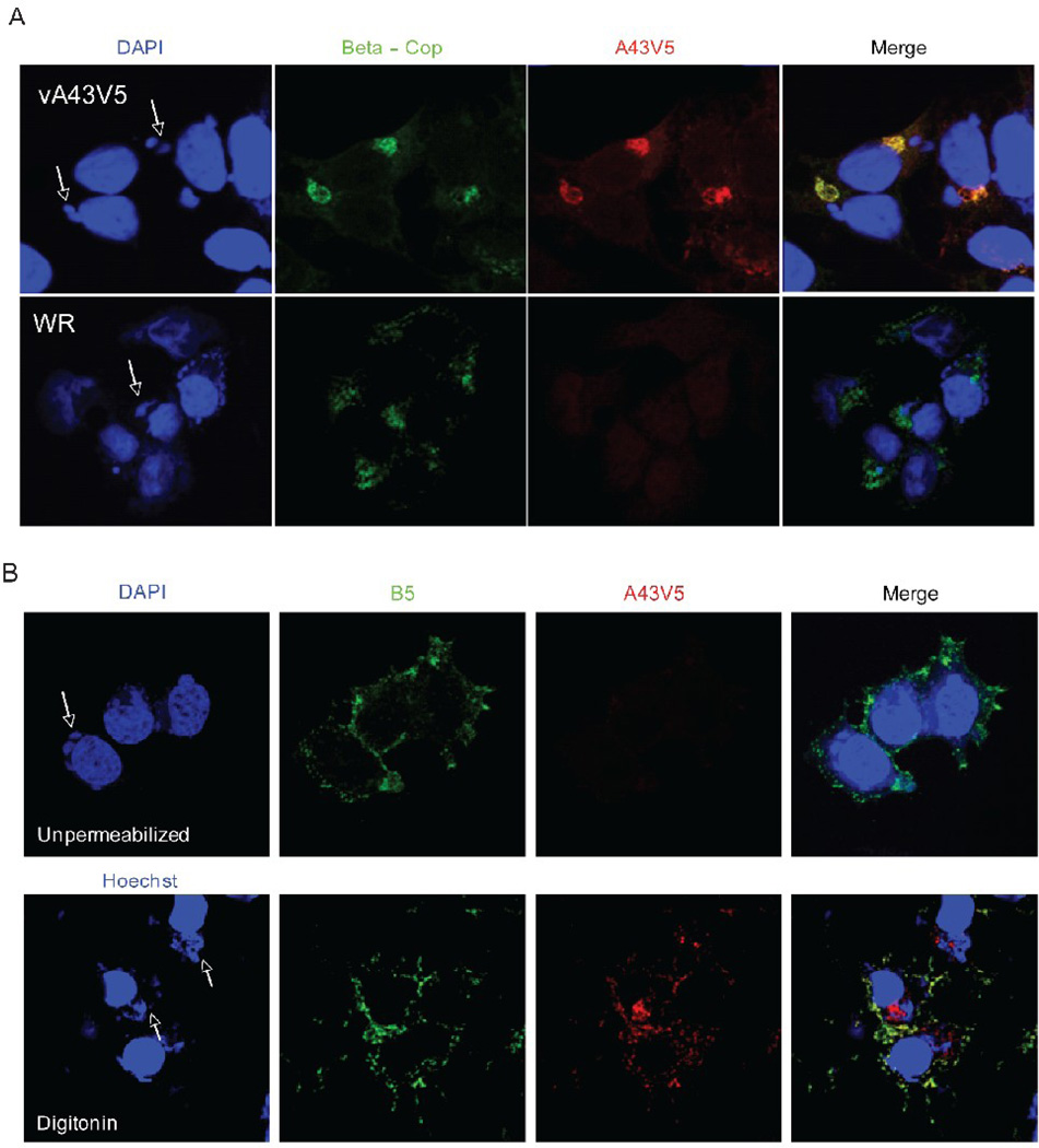
FIG. 5.
Intracellular localization and topology of A43. (A) Intracellular localization. HeLa cells were infected with VACV (WR) or vA43V5 at a multiplicity of 0.5 PFU per cell. At 24 h after infection, cells were fixed, permeabilized and stained with antibodies to V5 and β-Cop followed by fluorescently labeled secondary antibodies (red and green respectively). DNA was stained with DAPI (blue). Confocal microscopy images are shown. Arrows point to cytoplasmic virus factories. (B) Topology. Hela cells infected with vA43V5 were fixed and left unpermeabilized (top panels) or permeabilized with digitonin (bottom panels). Cells were then stained with an antibody to the EV protein B5 (green) as well as with an antibody to the V5 epitope tag (red) followed by fluorescently labeled secondary antibodies. DAPI or Hochest were used to stain DNA (blue). Confocal microscopy images are shown. Arrows point to cytoplasmic virus factories.

