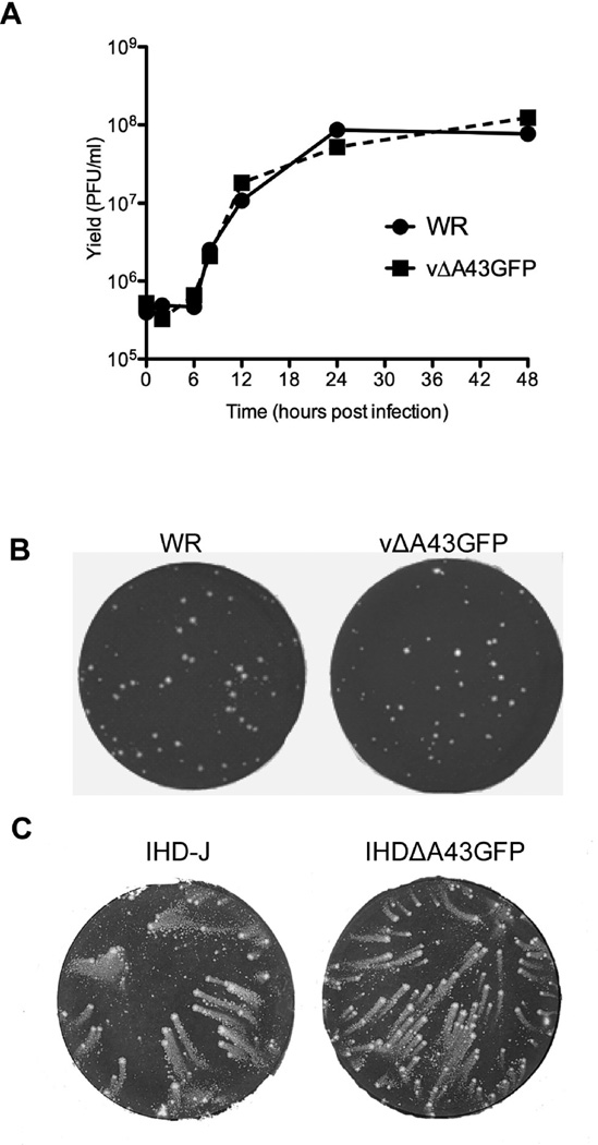
FIG. 7.
Replication of A43R deletion mutant. (A) One-step growth curve. BS-C-1 cells were infected at a multiplicity of 10 PFU/cell with vΔA43GFP and VACV WR. Virus yields were determined at the indicated times post infection by plaque assay. (B) Formation of vΔA43GFP and VACV WR plaques. BS-C-1 cells were infected with vΔA43GFP and VACV WR using a methylcellulose overlay. Cells were fixed and stained with crystal violet at 48 h after infection. (C) Comet formation. BS-C-1 cells were infected with VACV IHD-J or the A43 deletion mutant in the IHD-J background ΔA43GFP using a liquid overlay. After 48 h, the cells were stained as in panel A.

