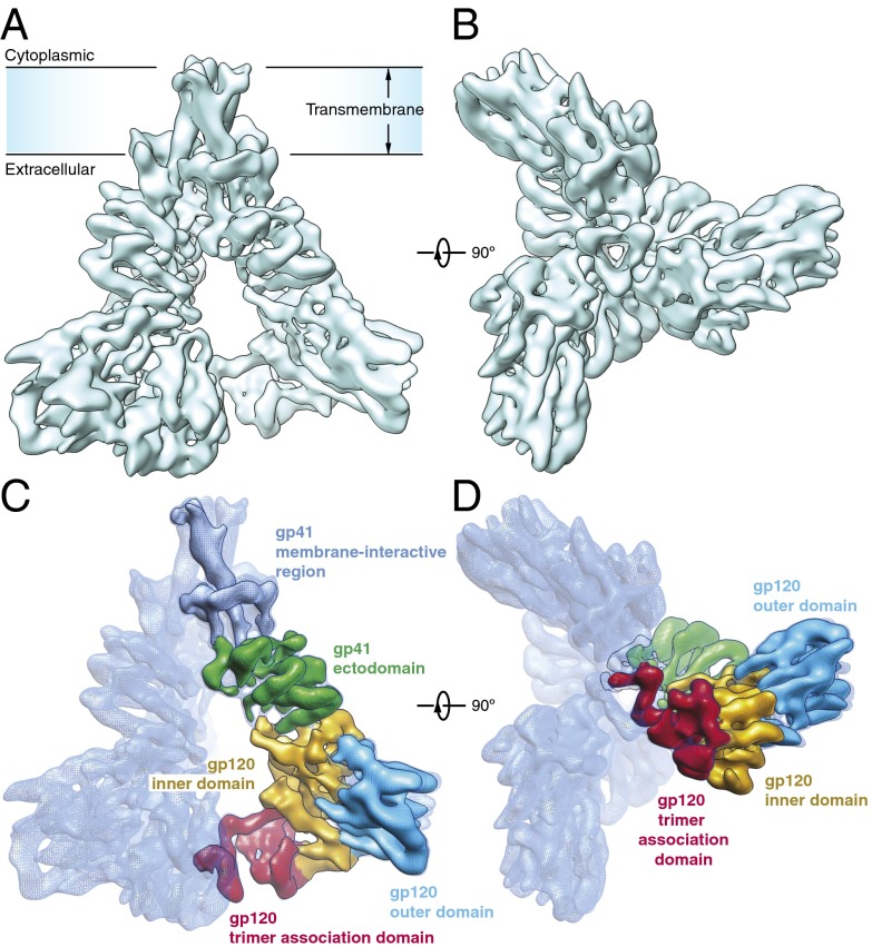
Fig. 1.
Architecture of the HIV-1 Env trimer. (A) Cryo-EM map of the HIV-1JR-FL Env trimer in a surface representation, viewed from a perspective parallel to the viral membrane. (B) Cryo-EM map of the HIV-1JR-FL Env trimer, viewed from the perspective of the target cell. (C and D) Domain organization of the Env protomer, revealed by segmentation of the density map. The gp120 domains are colored as follows: outer domain, blue; inner domain, orange; and TAD, red. The gp41 domains are colored as follows: ectodomain, green; and transmembrane region, cyan.

