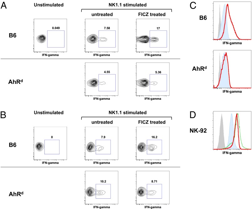
Fig. 4.
Activation of AhR enhances NK cell IFN-γ expression. (A) Splenocytes from WT and AhRd mice, injected with FICZ (3 μg per mouse) or vehicle control 48 h earlier, were stimulated ex vivo with plate-bound anti-NK1.1 antibody, stained for intracellular IFN-γ, and analyzed by FACS. Gate, DAPI−CD3−NKP46+. (B) Splenocytes (10 × 106) from WT and AhRd mice (Ly5.2) were adoptively transferred into B6 Ly5.1 mice. The mice were then treated with FICZ (3 μg per mouse) or vehicle control. The recipient spleens were harvested 72 h later and stimulated and analyzed as in A. Gate, Ly5.2 donor cells. (C) IL-2 (400 U/mL) cultured splenic NK cells from WT and AhRd mice were treated with FICZ (200 nM) for 7 h and stained for IFN-γ. Blue, IFN-γ staining; gray, unstained; red, IFN-γ staining in presence of FICZ. (D) Human NK-92MI NK cell line was treated with FICZ (200 nM) for 7 h and stained for IFN-γ. Blue, IFN-γ staining; gray, unstained; green, IFN-γ staining in presence of PMA (200 nM); red, IFN-γ staining in presence of FICZ. Results in this figure were reproduced at least once.

