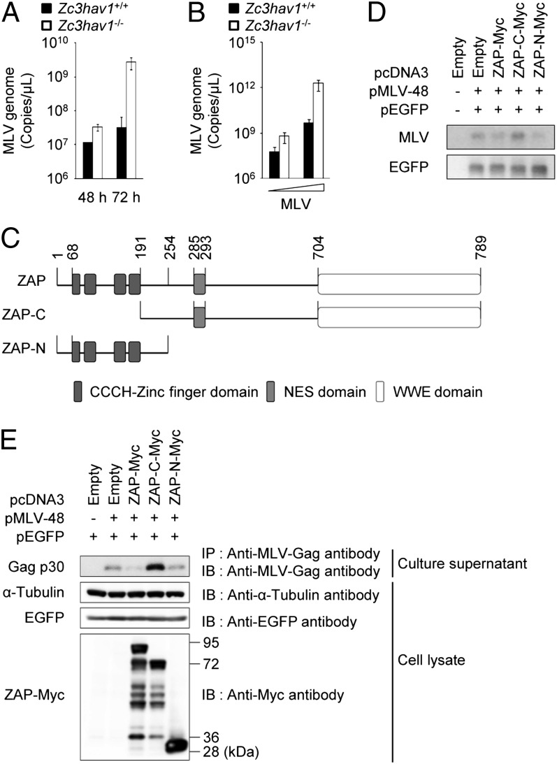
Fig. 2.
ZAP inhibits MLV replication in primary MEFs. (A) Zc3hav1+/+ and Zc3hav1−/− MEFs were infected with MLV (2 × 10¹⁰ copies per μL). Viral RNA was isolated at the indicated time points. The copy numbers of the MLV genome in the culture supernatants were measured by quantitative RT-PCR. (B) Zc3hav1+/+ and Zc3hav1−/− MEFs were infected with increasing doses of MLV (2 × 10⁸ and 2 × 10⁹ copies per μL) for 96 h. The copy numbers of the MLV genome in the culture supernatants were measured by quantitative RT-PCR. (C) Domain architecture of ZAP. (D and E) 293T cells were transfected with pMLV-48 and pEGFP-N1 together with the indicated ZAP expression plasmids for 48 h. Cytoplasmic RNA was subjected to Northern blotting analysis of the indicated RNAs (D). The culture supernatants were subjected to immunoprecipitation coupled to immunoblotting to detect the indicated proteins (E). The results shown are means ± SD (n = 3). NES, nuclear export signal.

