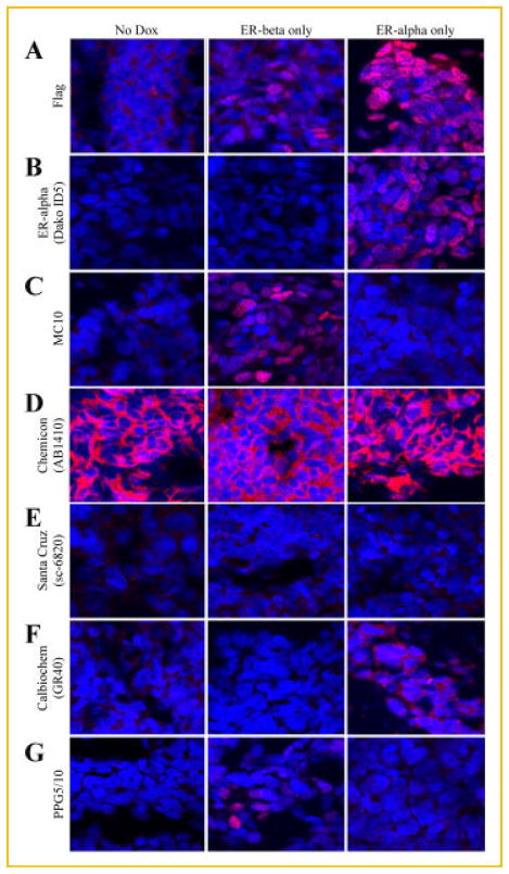
Fig. 4.
Specificity and comparison of the MC10 monoclonal antibody for ERβ protein to that of other commercial antibodies as determined by immunofluorescence. U2OS cells expressing either Flag-tagged ERβ or ERα under the control of a doxycycline inducible promoter were pelleted, paraffin embedded, sectioned, and processed for immunofluorescence. Sections of un-induced cells, ERβ expressing cells and ERα expressing cells were stained with a monoclonal Flag antibody (1:100) (A), an ERα antibody (Dako ID5, 1: 50) (B), or the following ERβ antibodies: MC10 (1:300) (C), Chemicon (AB1410, 1:400) (D), Santa Cruz (sc-6820, 1:100) (E), Calbiochem (GR40, 1:100) (F), and PPG5/10 (1:100) (G).

