
Figure 3.
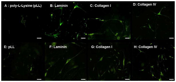
Neuronal differentiation on individual coated coverslips - βIII Tubulin antibody (green) was used to visualize neurons on Day 5 (A–D) and Day 15 (E–H) coverslips coated with pLL (A,E), Laminin (B,F), type I Collagen (C,G) and type IV Collagen (D,H). Enteric neurospheres on pLL barely initiate neuronal differentiation even at day 15. Neurospheres on Laminin, Collagen I and Collagen IV showed branching and several neuronal processes both at the early and late time points in vitro.