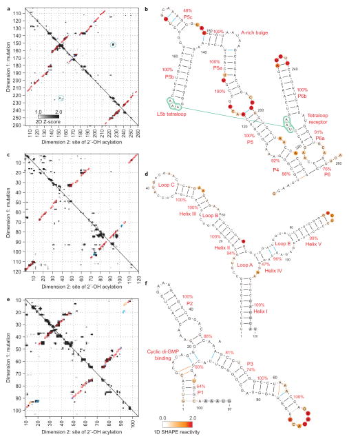
Figure 4. Accurate secondary structure models for non-coding RNAs.
a–f, Mutate-and-map Z-score data and resulting secondary structure models for the P4–P6 domain of the Tetrahymena group I ribozyme (a, b), the 5S ribosomal RNA from E. coli (c, d) and the domain that binds cyclic di-guanosine monophosphate from the V. cholerae VC1722 riboswitch (in the presence of 10 μM ligand; e, f). Colouring of squares (a, c, e) and lines and nucleotides (b, d, f) are as in Fig. 3.

