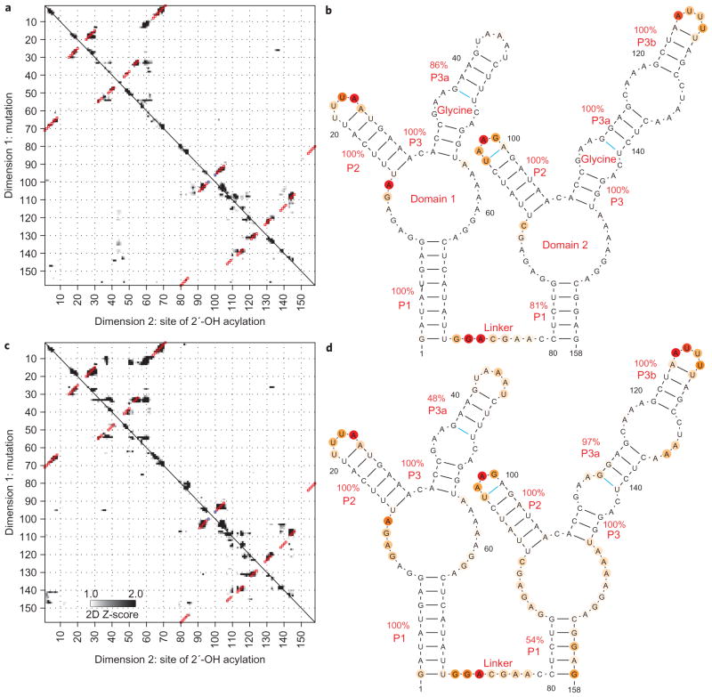
Figure 5. Two states of a glycine-binding riboswitch.
a–d, Mutate-and-map Z-score data and resulting secondary structure models for the double-ligand-binding domain of the F. nucleatum glycine riboswitch with 10 mM glycine (a, b) and without glycine (c, d), indicating no inter-domain helix swap upon glycine binding. Colouring of squares (a, c) and lines and nucleotides (b, d) are as in Fig. 3.

