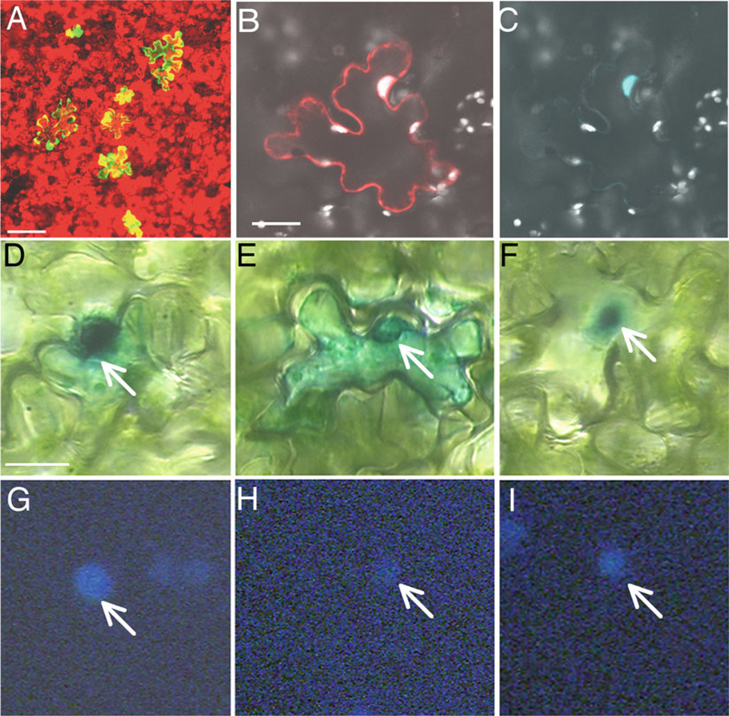
Fig. 1.
Examples of transient gene expression in leaf tissues following microbombardment. (a) Expression of a tandem repeat of the YFP gene in N. tabacum cv. Turk leaf (bar = 100 µ m), YFP expressing cells in yellow and chlorophyll auto fluorescence in red. (b, c) Expression of free DsRed and CFP-VirF fusion in a leaf cell of N. benthamiana (bar = 20 µ m), showing merged image with DsRed in red, CFP-VirF in blue, and chlorophyll autofluorescence in white (b) and CFP-VirF alone (c). For (a–c), observations were performed under a confocal microscope (Zeiss, LSM5 Pa), 24 h after microbombardment. (d–i) Localization of β-glucuronidase (GUS)-VirE2 fusion in A. thaliana leaves (bar = 10 β m). GUS-VirE2 is targeted to the nucleus in wild-type N. tabacum (d, g), whereas it is essentially cytoplasmic in vip1-antisense N. tabacum (e, h); in double transgenic vip1-antisense plants expressing VirE3, GUS-VirE2 nuclear localization is restored (f, i). Panels D–F represent GUS staining, and panels G–I represent DAPI staining. Arrows indicate cell nuclei. Histochemical GUS assay was done 24 h after microbombardment, and observation were performed after 3 h staining (as described in (13)).

