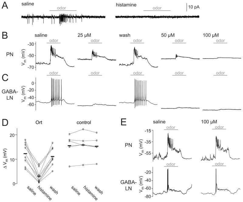
Figure 1. Histamine suppresses stimulus-evoked activity in Ort-expressing neurons.
(A) A cell-attached recording showing spikes in an Ort-expressing antennal lobe PN (left) in a regular saline bath. Subsequent bath application of histamine (100 μM) abolishes both spontaneous and odor-evoked spikes (right). Horizontal bars indicate the period (500 msec) of the odor stimulus. For all sample traces in this figure, the odor was pentyl acetate (10−2 dilution).
(B) A whole-cell recording of an Ort-expressing PN. Increasing concentrations of histamine produce greater suppression of activity, and suppression is reversed by wash-out. Note that this particular PN is depolarized by histamine, which we observed in several PNs; others were hyperpolarized or showed no change in resting membrane potential (see Discussion).
(C) Same as above, but in an Ort-expressing antennal lobe GABA-LN.
(D) Odor responses in saline, 100 μM histamine, and after wash-out (measured as the odor-evoked change in membrane potential). Each connected set of circles represents a different PN recording, and bars represent means (n = 8 Ort+ and 5 control). Histamine significantly reduces the odor response in Ort+ flies (p < 0.001, paired t-test). In control flies that lack the UAS-ort transgene, the odor response before and during histamine application is not significantly different (p = 0.69, paired t-test).
(E) Whole-cell recordings from a PN and a GABA-LN that did not express Ort. Histamine had no effect on these cells.

