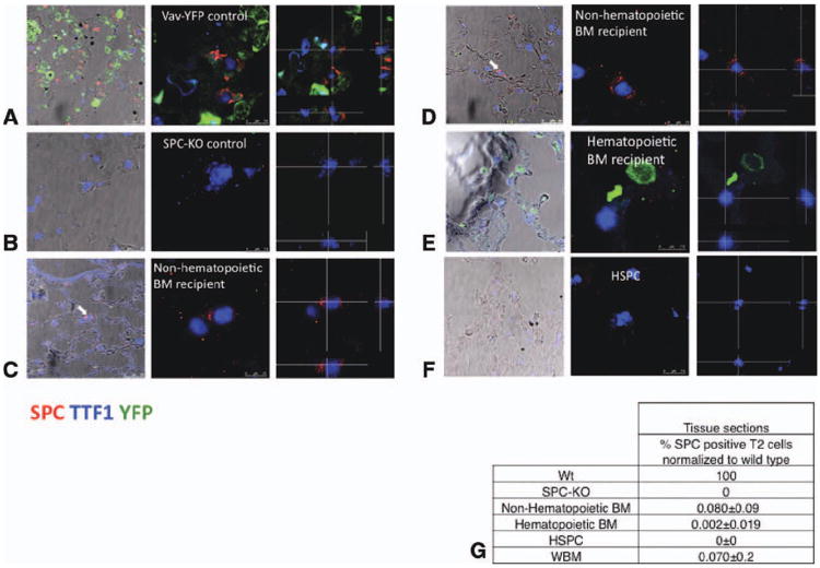
Figure 2. Analysis of lung tissue sections for donor-derived, SPC-positive cells.
(A)–(F): Confocal microscopic images of lung tissue sections stained for SPC, YFP, and TTF1. The left-most panels show images taken at low zoom, merged with bright-field images to show localization of type 2 cells within lung architecture. Donor-derived type 2 cells are marked with arrows. Middle panel: confocal images, ×630. Right panel: images were taken in 0.4-μm steps from the top to the bottom of each cell. Optical cross-sections through one focal plane show SPC expression in cytoplasmic vesicles surrounding the nucleus, excluding cell overlay. Cross-hairs indicate planes of optical x and y sections projected on bottom and right side. (A): Type 2 pneumocytes from vav-YFP control lungs coexpress vesicular SPC and nuclear TTF1. YFP is expressed in blood cells but not epithelial cells. (B): Type 2 pneumocytes in SPC-KO lungs express nuclear TTF1 but not SPC. (C) and (D): Lungs from two different SPC-KO mice that received nonhematopoietic BM cells contain donor-derived type 2 pneumocytes (vesicular cytoplasmic SPC and nuclear TTF1) located at the alveolar junction (arrows). (E): Lungs from SPC-KO mice transplanted with vav-YFP positive, hematopoietic BM cells contain vav-YFP positive blood cells. (F): Lung from SPC-KO mouse transplanted with wild-type HSPC. (G): The percentage of donor-derived, SPC-positive type 2 pneumocytes on tissue sections was calculated based on the percentage of type 2 pneumocytes among nucleated cells in corresponding wild-type samples, which was set to 100%. Abbreviations: BM, bone marrow; HSPC, hematopoietic stem and progenitor cell; TTF1, thyroid transcription factor 1; WBM, whole bone marrow; and Wt, wild type.

