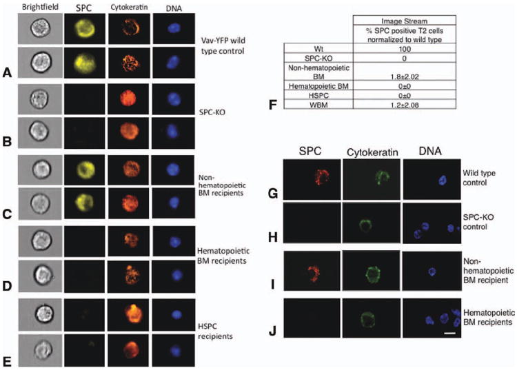
Figure 3. Detection of bone marrow-derived lung epithelial cells by ImageStream and confocal microscopy on single cells.
(A)–(E): Lung tissues from control (Wt and SPC-KO) mice or SPC-KO mice transplanted with the indicated donor BM population were digested with dispase, fixed, and analyzed for SPC (yellow), cytokeratin (orange), and YFP (green). Nuclei were stained with 4′,6-diamidino-2-phenylindole (DAPI; blue). Cells were analyzed using an ImageStream imaging flow cytometer with ×40 magnification (Amnis ImageStreamX). Examples of two cells from each group are shown. (A): Type 2 pneumocytes from a WT control mouse. (B): Lung cells from SPC-KO control mice. (C)–(E): Donor-derived, type 2 pneumocytes expressing SPC and cytokeratin were detected in SPC-KO mice transplanted with nonhematopoietic BM cells but not in mice receiving hematopoietic BM cells or purified HSPC. (F): The percentage of donor-derived, SPC-positive type 2 pneumocytes detected by ImageStream was calculated based on the number of type 2 pneumocytes among CD45-negative, round cells in corresponding wild-type samples, which was set to 100%. (G)–(J): Confocal microscopic analysis of sorted lung epithelial cells. Sorted cells were adhered to slides, fixed, and stained for SPC and cytokeratin. Nuclei were stained with DAPI. All cells shown were also CD45 negative (not shown). SPC-positive cells were detected in SPC-KO mice transplanted with nonhematopoietic BM cells but not in mice receiving hematopoietic BM cells. Scale bar = 7.5 μm is the same for all images. Abbreviations: BM, bone marrow; HSPC, hematopoietic stem and progenitor cell; WBM, whole bone marrow; and Wt, wild type.

