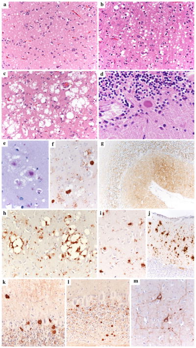
Fig. 2.

Distinctive histopathological features of sporadic human prion disease subtypes. a Typical spongiform change characterized by small, fine, microvacuoles (H&E stain, sCJD MM1, cerebral cortex); b spongiform change characterized by medium-sized vacuoles (H&E stain, sCJD VV1, cerebral cortex); c spongiform change characterized by relatively large confluent vacuoles (H&E stain, sCJD MM 2C cerebral cortex); d unicentric amyloid plaque of the kuru type (H&E stain, sCJD MV 2K, cerebellum); e, f florid plaques in the cerebral cortex (PAS stain and PrP IHC, vCJD, cerebral cortex); g synaptic pattern of PrP deposition in the cerebellum of sCJD MM1. A delicate diffuse staining is seen in the molecular layer, while the cerebellar glomeruli are stained in the granular cell layers. h Perivacuolar and i coarse PrPSc staining as typically seen in MM 2C and MM/MV 1+2C mixed sCJD cases. The cerebellum in MM 2C is either PrP negative or shows a focal patchy/coarse staining (j). k Plaque-like PrP deposits in the granular and molecular layers of the cerebellum in MV 2K. l Smaller plaque-like PrP deposits in the granular and molecular layers of the cerebellum in sCJD VV2. These plaque-like deposits are not visible in routine H&E or PAS-stained sections. m Perineuronal PrPSc staining in the deep cortical layers of sCJD VV2. All pictures in the panel have the same magnification (×200), except for d (×400) and g (×100)
