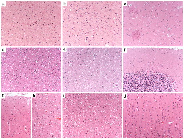
Fig. 3.
Distinctive regional distribution of histopathological changes in sCJD histotypes MM 2T, VV1, and VV2. Severe neuronal loss and gliosis in the medial thalamic (a) and inferior olivary nuclei (b) and relative sparing of anterior striatum (c) in sCJD MM 2T. Moderate to severe spongiform change (small to medium-sized vacuoles), gliosis, and neuronal loss in the cerebral cortex (d) and anterior striatum (e), and relative sparing of the cerebellum (f) in sCJD VV1; laminar distribution of spongiform change, with relative sparing of superficial (g) compared to the deep (h) layers in the cerebral neocortex of sCJD VV2. Severe spongiform change in the CA1/subiculum areas of the hippocampus (i) despite the virtually complete sparing of the occipital cortex (j) in a typical VV2 case with a relative short duration (five months). All pictures in the panel have the same magnification (×100) except for a and b (×200)

