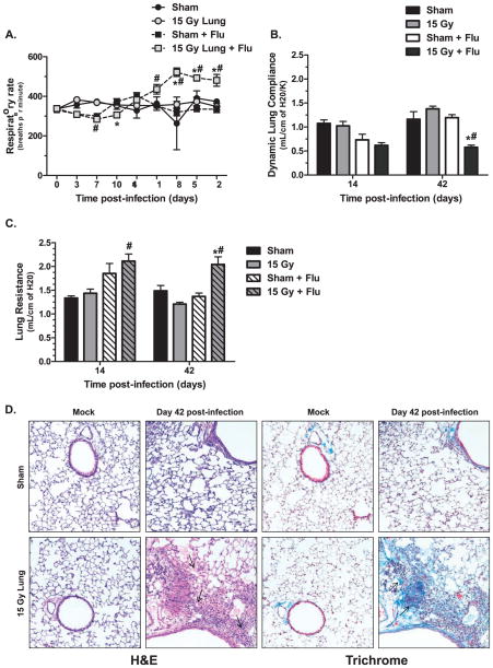
FIG. 2.
Lung function and pathology after infection in sham and 15 Gy whole-lung irradiated C57BL/6J mice. Whole-body plethysmography was used to assess pulmonary function changes after infection. Panel A: Respiratory rate over time. (*P < 0.05 versus mock-infected control, #P < 0.05 versus sham-infected, n =30 for virus-infected groups, n =7 for mock-infected groups.) Panel B: Dynamic lung complicance and (panel C) lung resistance at days 14 and 42 post-infection. Data are mean ± SEM of 3–6 animals per group. Data were analyzed by two-way ANOVA with Fisher’s post-test. (*P < 0.05 versus mock-infected control, #P < 0.05 versus sham-infected.) Panel D: Hematoxylin and eosin staining (H&E) (left two columns) and trichrome staining (right two columns) of sham and 15 Gy irradiated lungs at day 42 post mock or influenza A virus infection. Images (20×) are representative of 3–6 mice per group.

