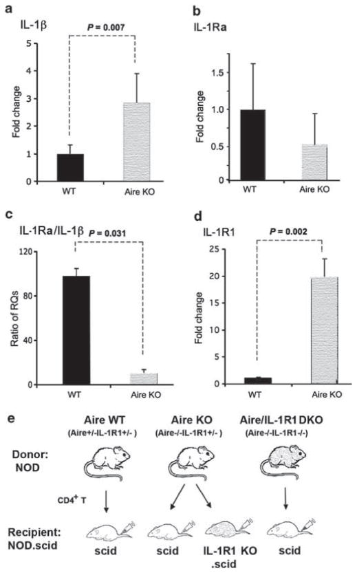
Figure 1.
Transcriptional profiling of ocular surface pro- and anti-inflammatory interleukin-1 (IL-1) cytokines in autoimmune regulator (Aire) wild-type (Aire WT) and Aire knockout (Aire KO) mice. Quantitative polymerase chain reaction (PCR) for (a) IL-1β, (b) IL-1 receptor antagonist (IL-1Ra), (c) IL-1Ra/IL-1β ratio and (d) IL-1R type 1 (IL-1R1). Expression in Aire WT controls was used as the reference (designated 1-fold) to generate relative quantitation (RQ) values. Data are shown as mean RQ value±s.d. In panel c, the ratio of IL-1Ra/IL-1β was obtained by dividing glyceraldehyde 3-phosphate dehydrogenase (GAPDH)-normalized RQ value of IL-1Ra by GAPDH-normalized RQ value of IL-1β in each mouse. Ratio data were shown as mean±s.d. Seven mice were studied per group. Unpaired t-test was used to test differences between groups; P<0.05 (indicated by dotted lines) was considered statistically significant. (e) Adoptive transfer design used to dissect the cellular mechanism of IL-1R1/IL-1 signaling in CD4+ T-cell-dependent autoimmunity.

