Figure 6.
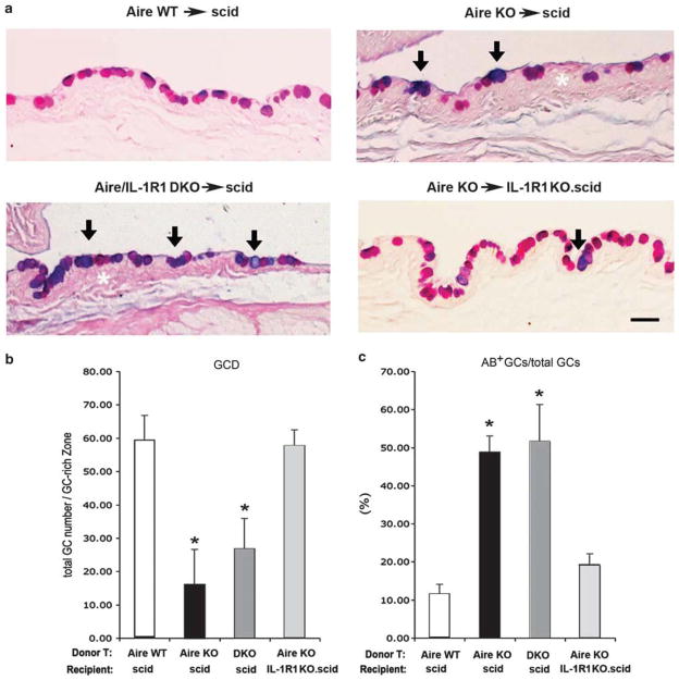
Histopathological evaluation of goblet cell density (GCD) and acidification by Alcian blue-periodic acid Schiff (AB-PAS) staining. (a) AB-PAS staining of the conjunctival GC-rich zone in each adoptive transfer group. GCs stained with both AB+ and PAS+ appeared dark blue/purple (arrows), in contrast to pink cells stained with PAS alone. White asterisks denote conjunctival epithelial hyperplasia in the GC-rich zone. Scale bar = 100 μm.
(b) Quantitative analysis of GCD as determined by the total GC number (sum of all AB- and PAS-stained cells) in a defined area of the GC-rich zone using × 100 magnification. Data are presented as mean±s.d. of total GC number/GC-rich area. (c) Percentage of AB+ cells in the total GC pool. Data are expressed as mean±s.d. of the percentage of AB+ GCs/total GCs. Asterisks indicate statistically significant differences vs the control group, Aire WT→scid, P<0.05. Aire WT, autoimmune regulator wild type; scid, severe-combined immunodeficiency.

