
Figure 2.
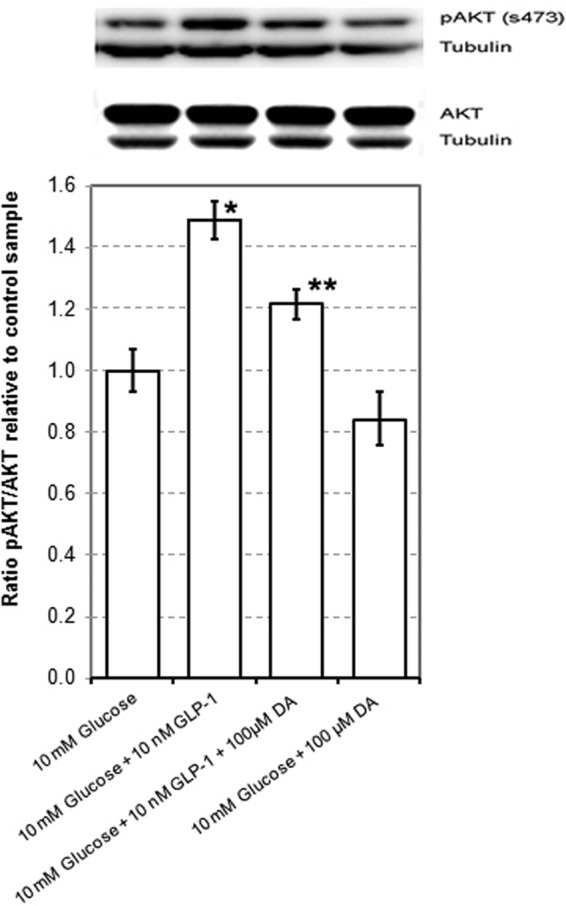
Dopamine Dampens GLP-1-Enhanced Glucose-Stimulated AKT Phosphorylation. INS-1E cells were grown to 80% confluence in 100-cm2 flasks, washed in PBS, and then rested for 1 hour in glucose-free RPMI supplemented with 2 mM glucose and 0.5% BSA. Cells were then incubated for 30 minutes in glucose-free RPMI containing 10 mM glucose, 10 nM GLP-1 and/or 100 μM dopamine. Protein lysates were prepared from cell monolayers, and equal amounts of protein were separated by reducing SDS-PAGE and analyzed by Western blot using anti-AKT and pAKT antibodies from CST. As a loading control, the amount of β-tubulin was also measured. The amount of immunoreactive protein on each blot was quantitated by the horseradish peroxidase-enhanced chemiluminescence reaction using a Flurochem M imaging station and associated imaging software. Western blot photograph from a representative experiment in a series of 4. The data from quantitation of immunodetected proteins are mean ± SE values from the same series of 4 experiments. The single asterisk denotes a statistically significant difference (P < .05) from the mean of 10 mM glucose control, and the double asterisk denotes a statistically significant difference from the 10 mM glucose plus GLP-1 group as determined by Student's t test. DA, dopamine.