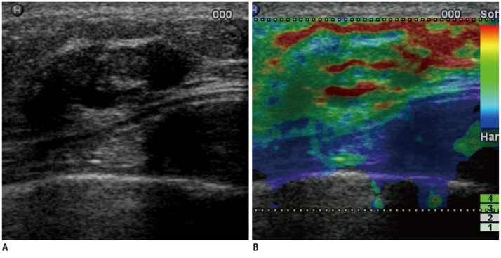
Fig. 2.
Forty nine-year-old woman with fibrocystic changes.
B-mode US image (A) shows oval, circumscribed, intracystic mass. Sonoelastographic image (B) shows entire solid component of complex mass as red or green, indicating soft lesion with elasticity score of 0. At B-mode US alone session, three of five readers classified lesion as likelihood of malignancy score of 2 (low suspicion for malignancy). Other two readers classified lesion as likelihood of malignancy score of 3 (intermediate suspicion for malignancy). For sonoelastographic image, all readers classified lesion as elasticity score of 0 (even strain for echogenic component). At B-mode US and sonoelastography session, all five readers downgraded lesion to likelihood of malignancy score of 1 (follow-up recommendation). US-guided core biopsy revealed fibrocystic changes. Lesion was stable during 2-year follow-up period. US = ultrasound

