Fig. 3.
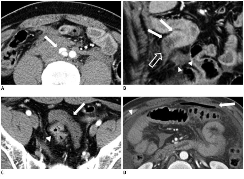
MDCT findings of jejunal segmental transection of 42-year-old man.
A. Axial contrast enhanced multidetector CT scan which was taken 1 hour after trauma shows focal dissection of right common iliac artery (arrow) and there is diffuse hematoma surrounding lesion. By focusing on this focal dissection, mesenteric hematoma around dissection was mistakenly thought to have originated from great vessel injury. B. Coronal reformatted contrast enhanced multidetector CT scan shows complete cut off of bowel loop at distal jejunum; complete cut off sign (open arrow). Jejunal loop shows abnormal dual bowel wall enhancement (both increased and decreased), and loses continuity soon after; Janus sign (arrows). Other end of broken bowel loop is identified nearby (arrowheads). C. Axial contrast enhanced multidetector CT scan presents 10 cm-long fragmented bowel segment in pelvic cavity (arrow). This fragmented bowel segment had no connection with adjacent bowel loop. There is no wall enhancing compared to normal enhanced bowel wall (arrowhead). D. Second axial contrast enhanced multidetector CT, taken 17 hours after trauma, shows free air (arrow) and increased amount of fluid (arrowhead) in abdomen. At first CT scans (A-C), there was no free air.

